The aim of this study was to estimate the subtle changes in cognitive processes assessed by the ERP technique under conditions where results of neuropsychological tests do not show any evident abnormalities. Additionally, it was intended to explore and verify the usefulness of n-back task in the ERP neurophysiological assessment of liver cirrhosis patients.
Subjects
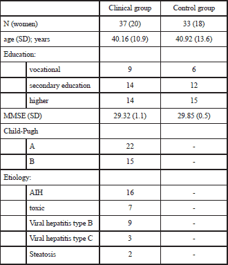
A clinical and control group were recruited among patients diagnosed and treated in the Gastroenterology and Hepatology Department of the University Hospital in Cracow. Patients with liver cirrhosis caused by viral infection (HBV and HCV) as well as autoimmune liver disease (autoimmune hepatitis (AIH), primary biliary cirrhosis (PBC), primary sclerosing cholangitis (PSC) and toxic liver cirrhosis (such as fat and medication) were included in the clinical group. The control group consisted of patients with functional gastrointestinal disorders such as irritable bowel syndrome (IBS) and functional dyspepsia. Subjects with a history of alcohol abuse or alcoholic liver cirrhosis were excluded because excessive alcohol use is an independent factor causing brain and cognitive abnormalities (8). Initially 138 patients were screened with the mini mental state examination (MMSE) for explicit cognitive abnormalities and those who scored below 27 points were rejected. Next, 109 patients were subjected to further neurological, neuroimaging and neuropsychological examination. In the neuropsychological assessment the normalized battery of tests was used. The scores below two standard deviations (S.D.) from the norm mean in at least two tests were considered as MHE. At these stages of selection, another 14 patients were excluded. In the following step, the data obtained from the ERP procedure were analyzed with regard to the task performance as well as technical and motor artifacts. Finally, data from 70 participants were the subject of further analysis. Table 1 show detailed demographic characteristics of the final group.
| Table 1. Demographic characteristics of subjects. Explanations: AIH - autoimmune hepatitis; MMSE - mini-mental state examination. |
 |
Neuropsychological tests
The PSE-syndrome-test or psychometric hepatic encephalopathy score (PHES) is the recommended psychometric battery for MHE assessment (4, 5). This battery consists of several simple paper-pencil tasks that are performed under time pressure. These tasks measure mainly attentional and graphomotor functions and have rather modest support in neuropsychological literature, except the trail making test (TMT) and digit symbol test. Recently, the International Society for Hepatic Encephalopathy and Nitrogen Metabolism (ISHEN) suggested the use of typical neuropsychological methods instead of PHES in order to more broadly assess cognitive functions (9). The measure of multiple cognitive domains has more clinical and diagnostic value than the use of simple graphomotor tests. In that way, MHE assessment was brought closer to cognitive assessment in other medical conditions. Due to the fact that the battery recommended by ISHEN is neither standardized nor normalized in Poland, the neuropsychological assessment was established by typical clinical methods, and the assessment lasted approximately 40 minutes. The battery contained: block design and digit symbol from the Polish adaptation of Wechsler adult intelligence scale - revised (WAIS-R) (10), TMT part A and B (11), the Polish versions of the verbal fluency test (VF) (12), auditory verbal learning test (AVLT) (13), and the computer version of the mental rotation test (MRT) (14).
The block design test requires manipulation of cubes in order to create a given pattern. It is intended to assess the level of constructional and visuo-spatial abilities. The digit symbol task requires the direct copying of simple graphics symbols, while the TMT part A requires the drawing of lines in order to sequentially connect numbers in a given order; it is performed as fast as possible and thus examines the speed of psychomotor responses as well as attention capacity. The TMT part B requires the connecting of numbers as well as letters in ascending order, alternating between the numbers and letters. It assesses the voluntary control of attention and efficiency of working memory, and can detect errors resulting from similar performance as in part A, e.g. connecting one kind of points: numbers or letters only. Lack of inhibition of prepotent responses is a symptom of the dysexecutive syndrome (e.g. in frontal and subcortical brain damage). The VF test requires the examinee to list as many words beginning with the letter "K" as possible (letter fluency) and name as many animals as possible (semantic fluency), both during a 1 minute period. This test assesses the efficiency of lexical search and access, ability to execute correct strategy for task performance and can detect perseverations and rule violations which suggest executive dysfunction. The AVLT is a standardized word-list learning task for episodic memory encoding and retrieval assessment as well as for examining the efficiency of verbal material retention and susceptibility to memory loss after a short distraction and a long delay. It can also examine the presence of perseverations and intrusions during learning and retrieval, which can be a sign of the dysexecutive syndrome. In the MRT test, subjects have to choose among four 3D rotated figures which are the same as the given target. It measures visuo-spatial abilities, but unlike the block design test, it is not related to motor performance and cannot be solved by applying trial-and-error strategy.
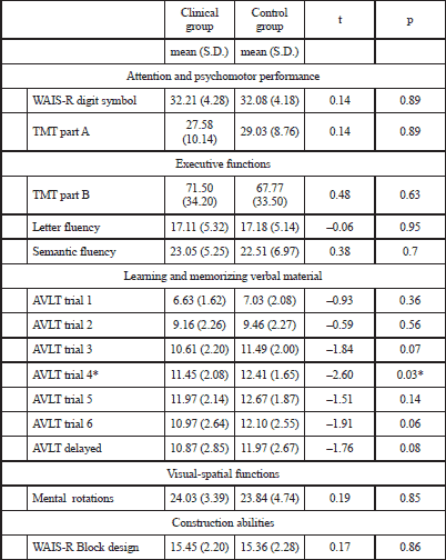
Results of the neuropsychological assessment are shown in Table 2. There are no differences between groups except the one for the learning trial of the AVLT. This single difference, however, has no clinical importance and is not sufficient for diagnosis of mild cognitive impairment or MHE. Moreover, this effect would diminish if additional correction for multiple comparisons had been used. We consider this effect as a statistical artifact due to the small sample size.
| Table 2. Neuropsychological test results. Asterisks indicate significant differences between groups (p<0.05; level not corrected for multiple comparisons). AVLT - auditory verbal learning test; TMT - trial making test; WAIS-R - Wechsler adult intelligence scale-revised. |
 |
N-back task
During the EEG investigation, the n-back task was administered. Series of letters were presented on a computer screen and the subject has to decide whether the letter currently shown is the same as shown n-places before. Two difficulty conditions (levels) were used. In the 1-back condition, the letter had to be compared to the previous one while in the 2-back condition to the letter shown two letters earlier. In both cases the YES/NO answer was selected by using computer keyboard. The procedure started with a short training session followed by the 1-back and then the 2-back condition. Each of the conditions consisted of 200 trials. In 33% of the trials, the letter shown was the same as the one presented 1- or 2-back earlier, respectively. During the presentation only capital letters were used (B, C, D, F, G, H, J, K, L, M, N, P, R, S, T, W, Z) with a presentation duration of 2 s. After the answer there was a fixation cross shown for 2 s. The cases in which the letter just shown was the same as the one to be compared with (which required a YES response) were called 'match'. Contrarily, the cases in which these letters were different (which required a NO response as the correct one) were called 'non-match' in further analysis.
All patients with very low behavioral performance were excluded from the EEG analysis. The d' parameter was applied as the exclusion criterion. It is a sensitivity index derived from the signal detection theory, commonly used in behavioral research on human perception and memory which allows to differentiate correct answers from the response bias. For calculating this index, only the 2-back task scores were taken into account, since it turned out to be difficult for all participants. The d' index was expected to reveal those participants who did not perform the task properly (e.g. they permanently selected the same answer in all trials, which led to either high false alarm or high miss rate), as well as low performance in performing the task. The average d' score was 0.89 (range: -0.97-1.87; S.D.: 0.57). The d' exclusion level was set to 0.2, which resulted in 11 participants being left out from further analyses.
Electroencephalography recording and analysis
For EEG recording, the Biosemi ActiveTwo device was used, equipped with 32 scalp Ag-AgCl active electrodes. They were mounted according to the 10-20 standard using EEG caps. Four additional electrodes over the eye muscles were used for off-line ocular artifact correction and two more for off-line linked mastoid reference calculation. EEG data were recorded with a sample rate of 256 Hz. After filtering in a range of 0.2-30 Hz, data were segmented into 1150 ms length bins (150 ms before and 1000 ms after stimulus onset). The segments were visually inspected in order to reject those containing recording artifacts. Next, ocular artifact correction was used according to the Gratton-Coles method (15) followed by remaining artifact rejection if max signal amplitude exceeded +/-30 µV in the given segment. Then, the segments related to correct responses were averaged separately for each condition.
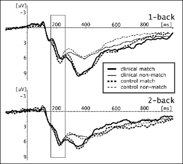
Visual inspection of grand averages revealed two time windows and two scalp areas for which EEG grand averages diverged between the groups. The first time window, 160-260 ms after the stimulus onset, was comprised of fronto-central derivations (F3, FC1, C3, Cz, Fz, C4, FC2, F4). The second one, 220-400 ms, was aggregated based on frontal electrodes (Fp1, AF3, F3, Fz, F4, AF4, Fp2). Grand averages are shown in Fig. 1 and Fig. 2.
 |
Fig. 1. ERP grand averages within the first time window 160-260 ms yielded from frontal-central derivations. |
 |
Fig. 2. ERP grand averages within the second time window 220-400 ms yielded from frontal derivations. |
Statistical analysis
The differences in neuropsychological tests between the groups were analyzed by parametric tests and p values less than 0.05 were considered as significant. Behavioral performance data from the n-back task as well as EEG data were analyzed using the 3-way repeated measures ANOVA with the difficulty condition (1- and 2-back) and correctness as within-subject factors and group (clinical and control) as a between-subject factor. In the behavioral analysis the dependent variable was the number of correct answers. In the EEG analysis, the averaged component amplitude within the considered time window was the dependent variable.
Both the clinical and control group had more correct responses in the 1-back task compared to the 2-back condition. They also had more correct responses in non-match than in match conditions.
The analysis revealed significant within-group main effects for congruence (F(1.68)=1307.08; p<0.001) and correctness (F(1.68)=11132.83; p<0.001) as well as for the interaction congruence correctness (F(1.68)=1018.55; p<0.001).
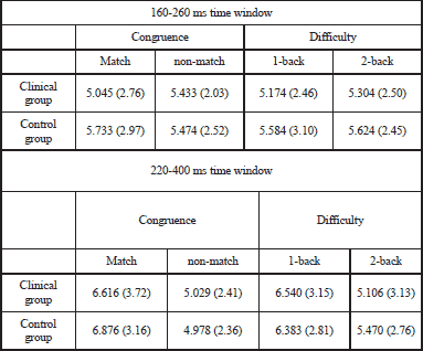
Mean ERP amplitudes for all considered conditions are shown in Table 3. In the first time window, there were no significant within-group main effects. However, statistical significance was reached by the interaction congruence x group (F(1.68)=4.33; p<0.05). In the second time window only within-group statistical effects were found: for difficulty (F(1.68)=4.72; p<0.05) and congruence (F(1.68)=2.10; p<0.001) as well as the interaction of these factors (F(1.68)=7.71; p<0.01). As expected, 'match' compared to 'non-match' condition was related to larger absolute amplitudes. This was also the case for 1-back compared to 2-back condition.
| Table 3. Mean ERP amplitudes within selected time windows (in µV). Mean values ±standard deviation (S.D.). |
 |
Most of the MHE studies using ERP methods reported so far have been devoted to a relatively simple "oddball" procedure where subjects have had to differentiate among two or three similar stimuli. This procedure can reveal a P3 ERP component. When delayed latency and decreased amplitude of P3 is observed, it suggests MHE in patients with liver cirrhosis without evident encephalopathy (16-18). Compared to typical neuropsychological tests, analysis of P3 parameters seems to be a better predictor of MHE. Moreover, it can be used for estimating the future course of the disease as well as its remission (7). On the other hand, analysis of a single P3 component in this task cannot reveal as much information concerning patients cognitive functioning as neuropsychological assessment does. This was our rationale for using a more sophisticated task which engages more diverse metal functions to a greater extent. The n-back task is a tool for working memory and executive functions assessment, so ERP results have a clinically useful behavioral context (16).
The expected behavioral effect in this task was a decrease in performance along with an increase in the n parameter. On the other hand, it was expected that the P3 ERP component would be attenuated in this situation (19, 20). In the present study both expectations were fulfilled. A decrease of ERP component amplitude was found in the second time window (220-440 ms), when the frontal P3 component could be observed. The lack of between-group differences in this range is not surprising, since the clinical group was comprised of patients with liver cirrhosis with neither MHE nor evident encephalopathy diagnosed.
Even though both groups did not differ according to task correctness, some subtle differences could be observed. This was visible in frontal electrodes in the time window of 160-260 ms, which could be considered as a P2 component. The functional significance of this component is less known. However, it is established that for visual stimuli, P2 amplitude correlates with the function of differentiating stimuli and directing attention to another task relevant stimulus (21). It is hypothesized that changes in P2 amplitude can be linked with changes in attention redirection related to appraisal of correctness/incorrectness of the given letter (22).
The results show that ERP methods can be useful in diagnosis of MHE since they show higher sensitivity in detecting subtle changes than the set of tests chosen in the present study. The relationship between observed group differences and cognitive functions measured by the tests is not clear. It is possible that the tests did not measure directly those attention functions which were correlated with the P2 component which differentiated between groups. Successful completion of a neuropsychological test requires many complex cognitive functions as well as a relatively longer time. In such tasks it is not possible to split them into very simple elementary activities such as moving the gaze onto the key stimulus or perceiving scene details. As a consequence, it is also impossible for the tests to assess cognitive activity related to activities which take no more than fractions of a second. On the other hand, the P2 component is related to such elementary perceptual and attention processing. ERP procedures are better suited to measuring such elementary processes.
The results of this study show that subtle alterations in brain functioning in liver cirrhosis can be detected even when patients do not fulfill MHE criteria. It is not clear whether such alterations are a prodromal expression of MHE or are MHE independent. Since the sample size in the present research was relatively small, this issue should be addressed in future research.
The ERP technique is often applied to group comparisons in experimental research but not to individual assessment in clinical settings. Due to the equipment and technical requirements and specialized research training for interpretation, this method is not available in a clinical practice. The neuropsychological assessment remains the basic method for MHE diagnosis. But interpretation of neurophysiological alterations visible in the ERP method is difficult to relate them to neuropsychological tests results. Contrary to the "odd-ball" task often used for the MHE assessment, the n-back applied here is related to the working memory and executive attention. These cognitive domains have clinical and practical importance as well as are theoretically supported. Moreover, assessment of working memory is also adopted in animal model of liver cirrhosis (23) and other cognitive impairment (24).
The results of this study show that n-back task is very sensitive in detection of subtle neurocognitive alterations in liver cirrhosis. This suggests its further exploration in future research as a method more consistent with other cognitive assessment.
Acknowledgments: The study was financially supported by the Polish Ministry of Science and Higher Education (grant no N N404 153134).
Conflict of interests: None declared.
- Guevara M, Baccaro ME, Gomez-Anson B, et al. Cerebral magnetic resonance imaging reveals marked abnormalities of brain tissue density in patients with cirrhosis without overt hepatic encephalopathy. J Hepatol 2011; 55: 564-573.
- Ilic S, Drmic D, Zarkovic K, et al. High hepatotoxic dose of paracetamol produces generalized convulsions and brain damage in rats. A counteraction with the stable gastric pentadecapeptide BPC 157 (PL 14736). J Physiol Pharmacol 2010; 61: 241-250.
- Rodrigo R, Felipo V. Brain regional alterations in the modulation of the glutamate-nitric oxide-cGMP pathway in liver cirrhosis. Role of hyperammonemia and cell types involved. Neurochem Int 2006; 48: 472-477.
- Dhiman RK, Saraswat VA, Sharma BK, et al. Minimal hepatic encephalopathy: consensus statement of a working party of the Indian National Association for Study of the Liver. J Gastroenterol Hepatol 2010; 25: 1029-1041.
- Ferenci P, Lockwood A, Mullen K, Tarter R, Weissenborn K, Blei AT. Hepatic encephalopathy - definition, nomenclature, diagnosis, and quantification: final report of the working party at the 11th World Congresses of Gastroenterology, Vienna, 1998. Hepatology 2002; 35: 716-721.
- Amodio P, Campagna F, Olianas S, et al. Detection of minimal hepatic encephalopathy: normalization and optimization of the psychometric hepatic encephalopathy score. A neuropsychological and quantified EEG study. J Hepatol 2008; 49: 346-353.
- Saxena N, Bhatia M, Yoshi Y, Garg P, Tandon R. Utility of P300 auditory event related potential in detecting cognitive dysfunction in patients with cirrhosis of the liver. Neurol India 2001; 49: 350-354.
- Fadda F, Rossetti ZL. Chronic ethanol consumption: from neuroadaptation to neurodegeneration. Prog Neurobiol 1998; 56: 385-431.
- Randolph C, Hilsabeck R, Kato A, et al. Neuropsychological assessment of hepatic encephalopathy: ISHEN practice guidelines. Liver Int 2009; 29: 629-635.
- Brzezinski J, Gaul M, Hornowska E, Jaworowska A, Machowski A, Zakrzewska M. Skala inteligencji Wechslera dla doroslych. Wersja zrewidowana - renormalizacja. Polska adaptacja WAIS-R (PL). Warszawa, Pracownia Testow Psychologicznych Polskiego Towarzystwa Psychologicznego, 2004.
- Strauss E, Sherman EMS, Spreen O. A compendium of neuropsychological tests: administration, norms, and commentary. New York, Oxford University Press, 2006.
- Lezak M, Howieson D, Loring D. Neuropsychological assessment. New York, Oxford University Press, 2004.
- Choynowski M, Kostro B. Podrecznik do Testu pietnastu slow. In: Testy psychologiczne w poradnictwie wychowawczo-zawodowym, M Choynowski (ed). Warszawa, PWN, 1977, pp. 102-169.
- Shepard RN, Metzler J. Mental rotation of three-dimensional objects. Science 1971; 171: 701-703.
- Gratton G, Coles MG, Donchin E. A new method for off-line removal of ocular artifact. Electroencephalogr Clin Neurophysiol 1983; 55: 468-484.
- Weissenborn K, Scholz M, Hinrichs H, Wiltfang J, Schmidt F, Kunkel H. Neurophysiological assessment of early hepatic encephalopathy. Electroencephalogr Clin Neurophysiol 1990; 75: 289-295.
- Ciecko-Michalska I, Senderecka M, Szewczyk J, et al. Event-related cerebral potentials for the diagnosis of subclinical hepatic encephalopathy in patients with liver cirrhosis. Adv Med Sci 2006; 51: 273-277.
- Hollerbach S, Kullmann F, Frund R, et al. Auditory event-related cerebral potentials (P300) in hepatic encephalopathy-topographic distribution and correlation with clinical and psychometric assessment. Hepatogastroenterology 1997; 44: 1002-1012.
- Watter S, Geffen GM, Geffen LB. The n-back as a dual-task: P300 morphology under divided attention. Psychophysiology 2001; 38: 998-1003.
- McEvoy LK, Smith ME, Gevins A. Dynamic cortical networks of verbal and spatial working memory: effects of memory load and task practice. Cereb Cortex 1998; 8: 563-574.
- Luck SJ. An introduction to te event-related potential technique. Cambridge, MIT Press, 2005.
- Peters J, Suchan B, Zhang Y, Daum I. Visuo-verbal interactions in working memory: Evidence from event-related potentials. Brain Res Cogn Brain Res 2005; 25: 406-415.
- Mendez M, Mendez-Lopez M, Lopez L, Aller MA, Arias J, Arias JL. Working memory impairment and reduced hippocampal and prefrontal cortex c-Fos expression in a rat model of cirrhosis. Physiol Behav 2008; 95: 302-307.
- Stasiak A, Mussur M, Unzeta M, Lazewska D, Kiec-Kononowicz K, Fogel WA. The central histamine level in rat model of vascular dementia. J Physiol Pharmacol 2011; 62: 549-558.