NEUROPHARMACOLOGICAL INVESTIGATION, ULTRA-HIGH
PERFORMANCE LIQUID CHROMATOGRAPHY ANALYSIS, AND IN SILICO STUDIES
OF PHYLA NODIFLORA
INTRODUCTION
Neurological diseases impose a huge burden on global health and this impact is increasing exponentially in low-middle socioeconomic countries. The pathophysiology of these disarrays include the progressive neuronal degeneration that might start during developmental stage and lasts throughout life (1). The common representatives of neurological disorders are behavioral impairments that disturb the quality of life precipitating the life-long disabilities throughout the world. Furthermore, these ailments impose a huge social, psychological and economic problem which continue to increase with aging (2).
Alzheimer’s disease (AD), a common neurodegenerative disorder, has affected around 24 million of the world’s populace and the prevalence is predicted to quadruple by 2050 (3). The pathophysiology involves the tau protein and b-amyloid tangles accumulation in brain resulting in the troubled functionality of synapses due to increased proinflammatory cytokines and oxidative stress (4). The momentous cognitive damage seen in AD is contributed by the cholinergic neurodegeneration in the nucleus basalis of Meynert, crucially involved in acquisition, consolidation and memory retrieval (5). Among the limited therapeutic options available for AD, anticholinesterases are more frequently used, as they elevate the acetylcholine (Ach) level in the synaptic cleft by blocking its enzymatic degradation. Unfortunately, the short half-lives of currently approved anticholinesterases and their complicated side effects stimulate the attention of researchers towards the alternative resources to treat this disease (6).
Plants comprise a variety of phytocompounds that play a crucial role in the neuroprotection against exaggerated oxidative stress and inflammation participating in progression of neurological disorders (7). Globally, 60% of the world’s population rely on plant-originated remedies due to their better affordability, convenient accessibility and noticeable safety profiles as compared to synthetic drugs, and the proportion is raising to 80% in economically emerging countries (8). The prevailing proportion of brain disorders are directing the researcher’s interest to explore the new therapeutic options from plants as these resources are extensively acknowledged for the discovery of alternative therapeutic options against a range of ailments.
Phyla nodiflora (P. nodiflora) is a fast growing wild herb belonging to Verbenacea family which is known for a broad range of pharmacological properties. In Bangladesh, it is traditionally used to cure neurological, gynecological and rheumatic problems (9). Its benefits to treat diarrhea, dysuria and micturition have also been broadly reported in the literature (10, 11). The herb has also been employed as a remedial choice by the populace of the Central America to alleviate various respiratory distresses (12, 13). Recently, a study has reported the antidiarrheal, bronchodilator and hypotensive effects of P. nodiflora (14). Additionally, its anxiolytic and anticonvulsant potentials have also been revealed previously (15), suggesting the significance of this herb in neurological disorders.
The current study includes the exploration of phytocomposition, antioxidative and memory-improving properties of crude methanolic extract of Phyla nodiflora (Pn.Cr) by a set of in vitro experimentation. The chronically treated rats with Pn.Cr were exposed to behavioral and biochemical assessment to evaluate any possible anxiety and amnesia ameliorating impact of this plant. Additionally, the docking studies were also incorporated to predict the probable interactivity of detected phytocompounds with acetylcholinesterase, for a better understanding of cognition-enhancing potential of P. nodiflora.
MATERIAL AND METHODS
All animal studies were performed after availing authorization from departmental ethical committee of BZU, Multan (09/PEC/2015) and were accomplished by following instructions of the “Institute of Laboratory Animal Resources” (ILAR), Commission on Life Sciences, National Research Council (NRC, 1996).
Chemicals
The chemicals and drugs of research grade were used for this study. Diazepam was procured from Roche Pharma, Switzerland while piracetam (P5295) and scopolamine hydrobromide trihydrate (S1875) were supplied by Sigma-Aldrich, St. Louis, MO, USA. These drugs were dispensed freshly on the day of the experiment.
Preparation of plant extract
The plant material was collected fresh and after authentication from expert botanist (voucher no: R.R. Stewart 607) was shade-dried. The dried plant was subjected to coarse grinding with subsequent maceration in 70% methanol (Roth, Karlsruhe, Germany) in a light-resistant amber glass bottle. The mixture was shaken intermittently and was passed through filtration process after a period of a week. The whole process of maceration, extraction and filtration was repeated twice. The yielded filtrates were combined and evaporated until the brownish extract was obtained which was stored at –20°C.
Total phenolic and flavonoid content determination
The total phenols were estimated through Folin-Ciocalteau method (16). In detail, 0.5 ml of plant extract or standard gallic acid (Merck, Darmstadt, Germany) solutions were mixed with 0.1 ml of 0.5 N Folin-Ciocalteau reagent (Scharlab S.L., Barcelona, Spain) followed by addition of 2.5 ml of sodium carbonate (Na2CO3) (Duksan Pure Chemicals Co., Ansan, South Korea). After the incubation period of 30 min, the mixture was tested for absorbance at 760 nm using spectrometer (IRMECO UV-VIS spectrometer, model U2020, Geesthacht, Germany). The total phenolic content in the extract was then determined by standard curve method, and expressed as mg gallic acid equivalents per gram of the extract.
The aluminum chloride method was used for total flavonoid content according to the method described (17). The 1 mg/ml of Pn.Cr was mixed with 1 ml of 10% AlCl3 (Thermofisher Sci., Waltham, MA, USA) The absorbance of mixture was noted at 415 nm after an incubation period of 30 min and outcomes were expressed as mg quercetin equivalent unit per gram of the extract.
Determination of antioxidant activity
1. 2,2-diphenyl-1-picrylhydrazyl (DPPH) method
DPPH method was used to examine the oxidative stress-combating potential of Pn.Cr by adopting the reported methodology (18). Different concentrations of plant extract were prepared and then 0.125 ml of DPPH (Tokyo Chemical Industry Co., Ltd., Tokyo, Japan) and 0.375 ml of methanol were added in 0.5 ml of each concentration. After that, each mixture was subjected to incubation for 30 min and optical density was evaluated at 517 nm in the presence of ascorbic acid as a positive control.
2. ABTS method
The radical scavenging activity of the extract was further assessed by already described method with slight modification (19). The aqueous solution containing 7 mmol/L of ABTS (Molekula, Munchen, Germany), and 2.45 mmol/L of potassium persulfate (Duksan Pure Chemicals Co., Ansan, South Korea) were prepared separately and incubated together in equal quantities for 16 h in darkness to prepare the reaction solution which is further diluted before use. For an assay in 96 well plate, 195 µl of this solution was mixed with 5 µl of plant extract solution or standard, ascorbic acid (Merck, Darmstadt, Germany) solution and incubated for few mins (5 – 7 mins) at room temperature. The absorbance was noted at 734 nm by using 95% of ethanol (Roth, Karlsruhe, Germany) as a blank by microplate reader (Spectramax 340PC384, Molecular Devices, USA). The free radical scavenging activity of plant extract was evaluated by following formula:
% radical scavenging activity =
(Abs control – Abs sample/Abs control) × 100
3. Anticholinesterase (AChE) activity
For assessment of AChE activity, 250 µl of Pn.Cr, 20 µl of DTNB (10 mM) (Molekula, Munchen, Germany) and 10 µl of 6.67 U/ml AChE (cat.#C3389, Electric EEL, Sigma-Aldrich, St. Louis, MO, USA) were mixed in 1710 µl of 50 mM Tris-HCL buffer (pH 8.0) (Merck, Darmstadt, Germany). After that, 10 µl of acetylthiocholine chloride (ATC) (200 mM) (Molekula, Munchen, Germany) was added to the reaction and absorbance was determined at 412 nm by employing galantamine (Reko Pharmacal, Lahore, Pakistan) as a positive control (20). Any change in absorbance was monitored for 5 min. The % inhibition of AChE was evaluated by using the following formula:
% inhibition = 100 – (Abs blank – Abs sample/Abs blank) × 100
Secondary metabolites analysis by ultra-high performance liquid chromatography-mass spectrometry (UHPLC-MS)
The Pn.Cr was examined for secondary metabolites through UHPLC-MS analysis. For this study, Agilent 1290 Infinity LC system along with Agilent 6520 Accurate-Mass Q-TOF mass spectrometer with dual ESI source (Agilent Technology Inc., Santa Clara, CA, USA) was used. The column used for UHPLC analysis was Agilent Zorbax Eclipse XDB-C18, narrow-bore 2.1 × 150 mm, 3.5 micron. The temperature was regulated at 25°C for column while at 4°C for auto-sampler. The 1.0 µL of sample was injected and its flow was maintained at 0.5 mL/min. Sample run time was 25 min and post-run time was 5 min. The mobile phases used in the analysis were formic acid (0.1%) (A) and in acetonitrile (B). Full scan MS analysis was done using negative electrospray ionization mode, over a range of m/z 100 – 1000. Nitrogen was supplied at flow rates of 25 and 600 L/hour as nebulizing and drying gas, respectively and the drying gas temperature was kept at 350°C. The capillary voltage was set to 3500 V while the fragmentation voltage was optimized to 125 V. Obtained data was examined through Agilent Mass Hunter software (B.05.00) for qualitative analysis (method: Metabolomics-2017-00004.m) and the detected compounds were identified through Search Database (METLIN_AM_PCDL-N- 170502.cdb).
Animals
The male Sprague Dawley (SD) rats (100 – 150 grams) of 4 – 6 weeks were used in this study, which were purchased from the National Institute of Health, Islamabad, Pakistan. The animals were accommodated separately in standard hygienic conditions maintained at 25 ± 2°C in the locality of Faculty of Pharmacy, B.Z.U, Multan. The rats were fed with customary rodent food containing 60% carbohydrates and 21% protein components with ad libitum approach to water. The rats were adapted to investigational handling for 2 – 3 days prior to experimental execution to circumvent the possibility of handling-induced stress.
The current study on animals was formally approved by the Departmental Ethical Committee of Bahauddin Zakariya University, Multan vide letter number 09/PEC/2015. All procedures were in accordance with the guidelines directed by Institute of Laboratory Animal Resources (ILAR), directive on Life Sciences, National Research Council (NRC, 1996).
Grouping of animals
Randomly selected animals were divided in six groups (n = 10) considering 1st group as control and treated with 1 ml/kg normal saline (0.9% w/v sodium chloride) only. Groups 2nd – 4th were designated as test animals and treated once orally with Pn.Cr 100 (Group 2nd), 200 (Group 3rd) and 300 mg/kg (Group 4th). Animals of the 1st – 4th group were treated with respective treatment for 30 consecutive days.
For the anxiolytic study, the 5th group was considered as positive control and treated with diazepam (2 mg/kg, i.p), 60 minutes prior to experiment. For learning and memory tests, the animals of the 5th group were administered with piracetam (200 mg/kg, i.p.) for seven days before the start of the y-maze. The 6th group was also added for the learning and memory test and was given scopolamine (1 mg/kg, i.p.) about 30 minutes before the start of each behavioral experiment for learning and memory. During learning and memory tests animals of all groups (group 2nd to 5th) except that of 1st group were given scopolamine (1 mg/kg, i.p.) 30 minutes before experiment, to precipitate the memory impairment and the outcomes were analyzed by comparing with the 6th group (scopolamine-treated amnesic rats). The details of animal grouping with respective treatments is given in Fig. 1.

Open field test (OFT)
This test is broadly applied to check the effect of test materials on animal’s anxiety and locomotor activity. It is a square-shaped (80 × 80 cm) open field made up of acrylic sheets and surrounded by 40 cm elevated walls (21). Before the start of the experiment, the open field arena was thoroughly cleaned with ethanol. On the day of the experiment, every animal was permitted to travel in the open field arena for 5 minutes. The animal’s preference for central zone was estimated by their entries and the duration of time spent in central zone through ANY-maze version 6.1, Stoelting Co. Wood Dale, JL, USA. Increased preference of central area of open field arena was the sign of reduced anxiety in the animal. Moreover, the total number of rearings and line crossings were also observed to evaluate the animal’s locomotion.
Light and dark box (L/D) test
The light and dark box is another alternative method for the analysis of the anxiolytic effect of the test material. This test uses the rodent’s behavior of reduced preference for illuminated areas as they get innately anxious towards such zones (22). The apparatus consists of two cabins made up of acrylic sheets. These cabins were designated as light and dark compartments and have dimensions of 40 × 40 cm and 40 × 33 cm, respectively. Both cabins are interconnected via a 10 × 10 cm door. Before experimentation, all animals were treated with designated treatment and subjected to behavioral testing one hour after the treatment. All animals were individually monitored for 5 minutes for their entries and duration of stay in light and dark compartments. Animal showing more preference for light compartment was considered lesser anxious (23).
Elevated plus maze test (EPM)
The neurobehavioral researchers broadly employ the EPM test for assessment of treatments effect on rodent’s anxiety. The rats tend to stay in closed zones as exposed and elevated areas are anxiogenic for them (24). This method uses the animal’s instinct struggle between discovering versus circumventing potentially risky areas. The test maze mounted at 50 cm from the base comprises two exposed and two walled arms connected by a central square (25). After 30 min of administration of designated treatment, every rat was put in the center and permitted to discover it for five minutes. The animal’s activity was captured through video recording and parameters such as an entry in the open arm and the duration of stay were monitored.
Behavioral test for memory and learning
1. Y-maze test
The test apparatus consists of three (A, B and C) arms (50 × 10 × 15 cm) connected at 120° to form a Y-shaped maze. This test assesses the short-term memory in rodents by estimating their spontaneous alternation behavior (26, 27). After 0.5 h of administration of group-wise chosen treatment, they were treated with scopolamine (1 mg/kg, i.p). Subsequently, 30 minutes after scopolamine treatment, every animal was tested for 5 minutes by monitoring the exploratory behavior in all three arms. The order of arm entries (ACB, CBA, BAC etc.) was noted for each animal to measure the spontaneous alternation (SAP). There is innate curiosity in rodents to explore the newer places and the animal having intact memory prefers the new arm over the visited one. The scopolamine administration results in amnesia and such animals reveal the least remembrance of the pre-explored arms and show decreased % SAP which was measured through following formula:
% Spontaneous alternation =
[number of alternations / total number of all arm entries -2] ×100
2. Novel object recognition (NOR) test
This testing paradigm evaluates the cognitive ability of rodents by exposing them to indifferent objects, as the rodents with efficient cognition are more inclined to explore the novel environment (17). On experiment day, the animals treated with designated treatment were injected with scopolamine (1 mg/kg, i.p). After 30 minutes, every rat was individually placed in an open arena (80 × 80 cm). Among two test sessions, rats were exposed to geometrically identical two objects for ten minutes (1st session). Subsequently, one object was replaced by novel one and animal was again exposed to arena for 5 minutes (2nd session). The interaction of animal with both objects was observed to estimate the discrimination index using the given formula:
DI = [(T2-T1) ÷ (T2 + T1)]
where T1 = duration of interaction with old object and T2 = Duration of interaction with new object.
An increase in the discrimination index is a sign of better working memory.
3. Morris water maze (MWM) test
The apparatus for the Morris water maze test comprised of a round plastic fiber container (150 cm × 50 cm) with a movable platform (10 × 10 cm) in it. Based on the four poles (N,S,W,E), the water-containing maze was divided into four quadrants and equipped with proximal and distal geometrical cues to help animals in navigation (28). The total duration of the test was six continuous days subdivided as two days for animal training (visible platform), three days for their testing (hidden platform) and one probe day (without platform). The position of the platform was maintained constant during all days. In the initial two days of training phase, each animal was individually trained for 4 trainings per day. Each training session had a duration of 2 minutes in which animals were allowed to navigate and locate the visible platform. In case of failure to find the platform, that animal was slightly directed towards platform and kept on it for 30 seconds. In the subsequent three test days, the rats had to find the submerged platform in water which was made murky by addition of non-toxic material (white poster ink) within 120 seconds and their escape latencies were monitored. During probe day, the rats were tested for 120 seconds in the tank without platform to evaluate their memory of the platform quadrant by noticing their number of entries and duration of swimming there. The videos were analyzed by using ANY-maze software version 6.1, Stoelting Co., Wood Dale, IL, USA.
Biochemical studies
Soon after the completion of MWM, the randomly chosen rats (n = 4) from every group were used for biochemical analysis. Animals were anesthetized with isoflurane (5% v/v) (Piramal Critical Care Inc., Bethlehem, PA, USA) and after decapitation, their brains were extracted. The brain homogenate was prepared by centrifuging the mixture of 4 ml of phosphate buffer (pH 7.4) (phosphate buffer tablets (Merck, Darmstadt, Germany) and 0.4 gram of isolated brain tissue for 20 minutes at 12,000 rpm under reduced temperature. The resulting supernatant was used to measure the enzymatic activities of by adopting the protocols described by (29).
Superoxide dismutase assay (SOD)
The 1 ml of Na2CO3 (50 mM), 0.2 ml of EDTA (0.1 mM) (Merck, Darmstadt, Germany), 0.4 ml of nitro blue tetrazolium (NBT) (Molekula, Munchen, Germany) were mixed with 0.5 ml of brain homogenate followed by the addition of 0.4 ml of hydroxylamine hydrochloride (HAC) (Merck, Darmstadt, Germany) and absorbance was checked at 570 nm (30).
Glutathione peroxidase assay (GPx)
The 200 µl of brain supernatant was mixed with 100 µl hydrogen peroxide (1 mM), (Sitara Chemical Industries Ltd., Faisalabad, Pakistan) 300 µl of 0.4 M phosphate buffer (pH 7.0), 200 µl reduced glutathione (2 mM) (Merck, Darmstadt, Germany) and 100 µl sodium azide (10 mM) (Merck, Darmstadt, Germany). After the incubation at 37°C for 15 min, 500 µl of 10% trichloroacetic acid (TCA) (Duksan Pure Chemicals Co., Ansan, South Korea) was added and mixture was centrifuged for 5 min at 1500 rpm. Later, 100 µl of this mixture was mixed with 200 µl of phosphate buffer (0.3 mM) and 700 µl of Ellman’s reagent (Molekula, Munchen, Germany) and absorbance was noted at 420 nm (31).
Malondialdehyde assay (MDA)
For MDA content estimation, the 3 ml of brain homogenate was mixed and boiled for 15 min with 1:1 proportion of trichloroacetic acid (TCA) and thiobarbituric acid (TBA) (Uni-Chem Co., Ltd., Wuxi-a, China). The mixture was later cooled and centrifuged at 3500 rpm for 10 min and absorbance was measured (32).
Acetylcholinesterase assay
Rats brain homogenate was also assayed for AChE activity by using a previously reported method (20). The 400 µl of prepared homogenate was mixed with 2.6 ml of 0.1 M phosphate buffer solution and 100 µl of DTNB was added to it. The absorbance of this control mixture was checked at 412 nm. Later, 5.2 µl of ATC was introduced into cuvette and change in absorbance was again noted for 10 min.
In silico studies
The affinity between detected phytocompounds of Pn.Cr and acetylcholinesterase was analyzed through molecular docking studies to evaluate their potential in targeting of acetylcholinesterase enzyme. The structure of target enzyme i.e. acetylcholinesterase in complex with donepezil (PDB ID:1EVE) was retrieved from RCSB databank (33). Structure based precision docking studies were carried out by setting a grid on the active site of AChE-donepezil interaction mode. Only chain A was retained for docking purposes, while the others, as well as the water molecules, binding inhibitors, and cofactors included inside, were removed by using Discovery Studio Visualizer (34). Polar hydrogens have been added to the obtained structure and saved as PDBQT format using AutoDock tools 1.5.4 for docking analysis (35). The validation of docking studies was carried out with the redocking of the donepezil into the active site of the prepared protein (1EVE-A). For that purposes, the structure of donepezil was prepared in PDBQT format and used as a reference in screening through AutoDock software with default parameters (35). The PDB structures for the ligands were prepared by using discovery studio. Energy-minimization and partial charges of the 3D structures of the studied phytocompounds were carried out with a convergence criterion of 0.01 kcal/mol. Å via SYBYL software through Tripos standard force field with Gasteiger-Huckel atomic partial charges according to Powell method (36). Then, AutoDock package was further used to prepare QTPDB formats of these phytocompounds which are needed for docking protocol. To cover the binding site in the protein, a grid box (x = 2.970, y = 63.635, z = 66.882 at 32 Å size) was generated by placing the donepezil as a center of the box. Finally, the docking of these phytocompounds was performed at the active binding site of the prepared protein and the interactions between the protein and ligands were evaluated using Autodock Vina set at default parameters (37) further analyzed by Discovery Studio Visualizer.
Statistical analysis
All data were first evaluated for normality by Kolmogorov-Smirnov test. All the behavioral and biochemical parameters were evaluated by the one-way ANOVA followed by post-hoc Dunnet’s test except, escape latencies and the distance travelled monitored during 3 days which were evaluated by two-way ANOVA followed by Tukey’s test. All the values were expressed as mean ± SEM and the results with P ≤ 0.05 were considered significant.
RESULTS
Total flavonoid and total phenolic content
The crude extract of Phyla nodiflora was evaluated for the estimation of total flavonoids and phenols. It was found that total flavonoids were 27.2 ± 4.3 mg QUE/g and total phenolic content was 79.4 ± 6.7 mg gallic acid/g in it (Table 1).

Determination of antioxidant activity
The anti-oxidant potential of plant extract was described by using ABTS and DPPH assays. The Pn.Cr showed dose-dependent antioxidant potential with IC50 values of 645.22 ± 23.5 µg/mL in ABTS assay while in DPPH assay it was 765.45 ± 25.41 µg/mL (Table 1).
Acetylcholinesterase inhibitory activity
Breakdown of acetylcholine may result in cognitive impairment and the drugs employed as therapeutic strategies in Alzheimer’s disease increases its levels. Anti-cholinesterase drugs inhibit the hydrolysis of acetylcholine and result in cognitive improvement. Therefore, acetylcholinesterase inhibitory activity of plant extract was evaluated by using Ellman’s method and concentration-based inhibition of enzyme was observed with IC50 value of 221.13 ± 12.41 µg/mL (Table 1).
Secondary metabolites analysis by UHPLC-MS
The Pn.Cr was subjected to ultra-high performance liquid chromatography mass spectrometry (UHPLC-MS) analysis to obtain a detailed secondary metabolites profile. The outcomes revealed the tentative existence of twenty-two phytocompounds as shown in Fig. 2 and Table 2. Most of these phytocompounds belonged to flavonoids and alkaloids. Meticulously, the ones from flavonoids were 3,4,7-trihydroxy-5-methoxy-8-prenylflavan 4-O-(beta-D-xylopyranosyl-(1->6)-beta-D-glucopyranoside), (-)-8-(2-carboxy-1-phenylethyl)-3,5,7-trihydroxyflavone delta-lactone, matteucinol 7-O-glucoside, kaempferol 3-(2G-glucosylrutinoside), isovitexin 7-(6’’’-sinapoylglucoside) 4’-glucoside, and 8-methoxycirsilineol. Whereas, the alkaloidal derivatives tentatively identified as per library database included mukoenine B, usambarensine, aromoline, fruticosonine, and delcosine. The remaining phytocompounds belonged to terpene (plocamene D and melledonol), acetogenin (laurenenyne A), chalcone (epoxyobovatachalcone), plant hormone (gibberellin A39), and dipeptide (hypoglycin B) groups of secondary metabolites.


Behavioral tests for anxiety
1. Open field test
After exposure of animals to open field, a noticeable intergroup variation for time spent at the center zone (F (4, 36) = 22.96, P < 0.0001) and the number of entries in center zone (F (4, 36) = 12.03, P < 0.0001) was observed. In brief, the animals treated with diazepam showed anxiolytic behavior by prominently increasing the preference for the central zone as compared to the rats in control group (P < 0.0001). Similarly, increased stay at the center zone was observed in the animals treated with Pn.Cr at 200 and 300 mg/kg as compared to control group animals which depicted a dose-dependent anxiolytic effect of the extract (P ≤ 0.005) (Fig. 3A). The number of entries in the central zone were also increased by the rats treated with Pn.Cr 200 mg/kg (P < 0.05) and Pn.Cr 300 mg/kg (P < 0.001) (Fig. 3B). The effect of extract treatment on animal’s locomotion was also monitored by evaluating number of line crossings and total rearings in rats. The one-way ANOVA represented that significant variation among the groups in line crossings (F (4, 36) = 20.69, P < 0.0001) and number of rearings (F (4, 36) = 27.45, P < 0.0001). The line crossings were increased in Pn.Cr-treated animals at dose 300 mg/kg (P < 0.01) while total rearings were increased at 200 mg (P < 0.05) and 300 mg/kg (P < 0.001) with respect to control (Fig. 3C and 3D). But, Pn.Cr at the dose of 100 mg/Kg did not show any significant outcomes in OFT.

2. Light and dark box test
The outcomes of L/D test showed that animals treated with Pn.Cr resulted in the reduction of anxiety as evidenced by the increased time spent in the light zone and decreased time spent in the dark zone in comparison with control (Fig. 4A and 4B). The outcomes of one-way ANOVA revealed intergroup difference in time spent in light box (F(4,35) = 40.2, P < 0.0001) and time spent in dark box (F(4,35) = 10.34, P < 0.0001). In brief, Dunnet’s multiple comparison test showed a significant difference for time spent in the light zone dose-dependently as Pn.Cr at 100, 200 and 300 mg/kg caused increased duration of stay in lightened zone as compared to control rats with P < 0.001 and P < 0.0001, respectively.

3. Elevated plus maze test
For the evaluation of anxiolytic effect of Pn.Cr, animals were exposed to open and closed arms in paradigm of elevated plus maze. The results showed that significant variation existed between differently treated animals for their number of entries in open arm (F(4,35) = 10.78, P < 0.0001) and duration of stay (F(4,35) = 10.3, P < 0.0001) there. The multiple comparison showed that Pn.Cr at 200 and 300 mg/kg significantly decreased the anxiety by increasing animal entries in the open arms with respective P < 0.001 and P < 0.0001 (Fig. 5A) and duration of stay there (Fig. 5B) with P < 0.01 and P < 0.001, as compared control animals.

Behavioral tests for memory and learning
1. Y-Maze test
The effect of Pn.Cr on spontaneous alternation in scopolamine-treated amnesic rats was studied by utilizing the Y-maze. The one-way ANOVA showed significance intergroup variation in %SAP (F(5,42) = 20.68, P < 0.0001) (Fig. 6). The administration of scopolamine resulted in the reduction of memory in rats as observed by a decrease in spontaneous alternation score in comparison to control group P < 0.0001. While administration of piracetam decreased the amnesic effect of scopolamine by increasing the %SAP (P < 0.0001) as compared with the scopolamine-treated group. Similarly, administration of Pn.Cr 200 and 300 mg/kg resulted in a dose-dependent increase in %SAP score with P < 0.05 and P < 0.0001, respectively.
 |
Fig. 6. The dose-dependent memory and learning affecting capacity of Pn.Cr was evaluated to reveal the animal’s % spontaneous alternation behavior using Y-maze. Data was analyzed by using one-way ANOVA followed by Dunnet’s test and shown as mean ± SEM. *P < 0.05 showing the significant difference for scopolamine-treated rats as compared to control. #P < 0.05 showing the significant difference for piracetam-treated rats as compared to the scopolamine-treated group. bP < 0.05 and cP < 0.05 were considered significant for Pn.Cr 200 and 300 mg/kg as compared to the scopolamine-treated group. |
2. Novel object recognition test
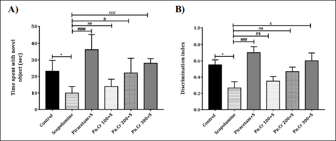
The rodents have the instinct capability to show more inclination towards the novel object if they have intact memory to remember the older object. The treatment of rats with scopolamine resulted in the development of amnesia as observed that scopolamine-treated animals spent lesser time with the new object as compared to control P < 0.05. Piracetam administration resulted in the prominent reversal of this scopolamine-induced memory loss (P < 0.0001). Likewise, the administration of Pn.Cr 200 and 300 mg/kg resulted in improvement in memory as observed by an increase in the time spent by these rats with the novel object with P < 0.05 and P < 0.001, respectively (Fig. 7A). However, Pn.Cr 100 mg/kg did not show any significant results. Discrimination index of Pn.Cr 100 and 200 mg/kg treated animals were not significantly different but piracetam and Pn.Cr 300 mg/kg showed significant results with respective P < 0.001 and P < 0.05 (Fig. 7B).
3. Morris water maze test
In MWM, the animals were trained to locate the hidden platform to examine the impact of Pn.Cr on spatial learning. It was observed that control group animals successfully searched out the platform during three consecutive days. Scopolamine-induced amnesia was observed by an increase in escape latencies, in comparison with the control group (F(1,100) = 188, P < 0.001). The treatment of animals with piracetam resulted in a decrease in escape latency and reversal of memory in comparison with the scopolamine-amnesic group (F(2,143) = 179.4, P < 0.0001). Also, the treatment of animals with plant extracts decreased the escape latencies as compared to control in a dose-dependent way (Fig. 8A).

Moreover, the scopolamine-treated animals traveled a long distance to find out the platform as compared to the control group (P < 0.0001). But, Pn.Cr treated animals showed a dose-dependent decrease in distance traveled to find the platform as compared to scopolamine-amnesic rats. The animals treated with 100, 200 and 300 mg/kg of Pn.Cr were capable of finding the platform in shorter time with P < 0.001, P < 0.001 and P < 0.0001, respectively as shown in Fig. 8B.
On probe day, the scopolamine-induced amnesia was evident form the reduced visits (P < 0.01) and swimming time (P < 0.05) in target quadrant as compared to control group. But, piracetam and Pn.Cr treated animals showed a dose-dependent improvement in remembrance of platform quadrant as 300 mg/kg caused increase in overall number of entries and time of swimming in the target zone (P < 0.001) (Fig. 8C and 8D).
Biochemical assays
The results of one-way ANOVA showed a significant difference among all differently treated groups (F(5,42) = 7.684, P < 0.0001). In detail, Dunnet’s test showed the elevation of acetylcholinesterase in scopolamine-treated rats as compared to control (P < 0.0001). The administration of piracetam prominently normalized the level of this enzyme with P < 0.001. Similarly, the concentration-dependent inhibition of acetylcholinesterase was noticed by 100, 200 and 300 mg/kg of Pn.Cr with respective P < 0.05, P < 0.001 and P < 0.0001, as compared to scopolamine-treated rats (Fig. 9A).

Superoxide dismutase combats the oxidative species and protects against the diseases induced by increased oxidative stress. T he results of biochemical enzyme activity exposed that significant intergroup variation occurs (F(5,42) = 41.26, P < 0.0001) for the level of superoxide dismutase. The enzyme levels were reduced in scopolamine-treated animals as compared to control (P < 0.01). But the treatment of animals with piracetam (P < 0.0001) and Pn.Cr 300 mg/kg (P < 0.001) resulted in a significant enhancement of enzyme level (Fig. 9B).
Glutathione is another defense mechanism of the body that provides protection against destructive peroxides. The One-way ANOVA analysis showed significant intergroup variation in the glutathione levels from the isolated brain homogenates (F(5,42) = 41.26, P < 0.0001). Briefly, the treatment of animals with scopolamine caused in a decrease in glutathione peroxidase as compared to control P < 0.0001. But piracetam reversed this loss prominently, as compared to scopolamine-treated rats with P = 0.0001. Similarly, Pn.Cr when administered to the animals resulted in a dose-dependent increase in enzyme levels at dose of 100 (P < 0.05), 200 (P < 0.05) and 300 mg/kg (P < 0.0001) (Fig. 9C).
The level of malonaldehyde (MDA) increases due to oxidative stress caused by the super-oxidation of lipids. The results unveiled the significant intergroup variation (F(5,42) = 18.6, P < 0.0001). The treatment of animals with scopolamine resulted in an elevation of its level in comparison with control animals (P < 0.0001). The treatment with piracetam reduced the enzyme level with respect to scopolamine-amnesic rats (P = 0.0001). Similarly, administration of Pn.Cr at 200 and 300 mg/kg resulted in the reduction of MDA levels with P < 0.05 and P < 0.0001, respectively (Fig. 9D).
In silico studies
In silico molecular docking technique was utilized to predict possible binding energy and modes of interactions of 22 compounds extracted from crude extract of Phyla nodiflora (Pn.Cr) in the active site of the 1EVE protein by the AutoDock Vina software. Estimated binding energies for 22 docked compounds are summarized in Table 3.

Based on these docking results, the top ten compounds were further evaluated for in silico pharmacokinetics parameters by using pkCSM (38) to analyze the potential of these compounds as possible drug candidates for future studies. For most of the drug candidates focusing on oral route of administration, high solubility favors complete absorption while the poor solubility limits the absorption of the drug in the gastrointestinal tract (Table 4).

We used pkCSM analysis to predict human intestinal absorption (HIA), blood-brain barrier penetration (BBB) and central nervous system (CNS) permeability.
Our pkCSM analysis showed that (-)-8-(2-carboxy-1-phenylethyl)-3,5,7-trihydroxyflavone delta-lactone, Epoxyobo-vatachalcone, Mukoenine B, Usambarensine, Glabrin D and Fruticosonine are able to penetrate the blood-brain barrier moderately. Additionally, log BB for the compound 12 is greater than 0.3, so it can be predicted that this compound is able to pass through the BB barrier readily. Usambarensine, appeared as the most promising ligand among the 4 proposed compounds, with the lowest binding energies of –12.00 Kcal/mol for the best interacting pose. There are three hydrogen bonds (HB) formed with the three amino acids (a conventional HB with TYR121 at a distance of 2.29 Å and two Pi-Donor HBs with SER122 and TYR334 at distances of 4.00 Å and 4.19 Å, respectively). The two Pi-Anion interactions with ASP72 at distances 3.11 Å and 3.46 Å and one Pi-Alkyl with PHE331 at distance 4.27 Å also minimize the conformational energy of the ligand. Usambarensine high affinity was also associated with the presence of two Pi-Pi Stacked and two Pi-Pi T-Shaped interactions with TYR334 and TRP279 (Fig. 10).

Another phytocompound, (-)-8-(2-carboxy-1-phenylethyl)-3,5,7-trihydroxyflavone delta-lactone binds with 1EVE protein via multiple hydrogen bonding and hydrophobic contacts. It could be observed from the Fig. 11 that they are three hydrogen bonds interactions with three different amino acids TRP84, TYR121 and TYR130 at distances of 2.44 Å, 2.99 Å and 3.34 Å, respectively and two Pi-donor interactions with TYR121 and SER122 at distance 3.10 Å and 3.78 Å, respectively. Further enquiry indicates the presence of Pi-Pi T-shaped interactions with TYR121 at distance 5.49 Å and PHE331 at distance of 5.75 Å. Presence of two Pi-Pi Stacked interactions with PHE331 and TYR334 at distances 5.15 Å and 3.91 Å, respectively was also observed (Fig. 11).

Epoxyobovatachalcone also showed good binding ability with acetylcholinesterase 1EVE having a docking score of –11.60 Kcal/mol for the best interaction pose. It forms a hydrogen bond with the amino acid TYR121 of the 1EVE protein at distance of 3.21 Å. Two hydrophobic interactions (Pi-Sigma with PHE331 at distance of 3.95 Å, three Pi-Pi Stacked two with TRP84 at distances 3.91 Å and 4.39 Å and one TYR334 at distance of 4.04 Å) minimize the conformational energy of the ligand. As these interactions are largely involve in charge transfer that helps in intercalating the ligand in the binding site of the protein (Fig. 12).

In silico studies also revealed good inhibiting interaction between Fruticosonine and acetylcholinesterase protein with docking score of –10.50 Kcal/mol for best interaction pose. Docking studies showed two hydrogen bonds formed between fruitcosonine and protein amino acids TYR121 and PHE288 at distance of 3.21 Å and 2.89 Å, respectively. Three additional interactions (Two Pi-alkyl interactions with TRP84 at distances of 3.73 Å and 4.03 5.25 Å and one Pi-Sigma with TYR334 at distance of 3.96 Å) also facilitate this interaction and minimize the conformational energy of the ligand (Fig. 13).

DISCUSSION
The disease-induced disabilities and mortalities are dominantly contributed by neurological disorders, worldwide. Among various brain disorders, dementia has affected approximately 50 million of the world’s population till 2020, 65% of which is attributed to AD. Due to the limited therapeutic options available to deal with this disorder and the resultant financial burden, the researchers are keen to discover the easily accessible and economic remedial entities with better safety profiles. Plants have been relied upon by almost 80% population in developing countries for the treatment of various ailments (39) as their phytoconstituents possess remedial properties beneficial for various ailments (40, 41).
In the light of previously reported literature on the medicinal benefits of Phyla nodiflora, the current study was planned to scientifically validate its neuropharmacological properties. The in vitro experiments on Pn.Cr was carried out to detect its polyphenolic content. Moreover, the detailed phytochemical composition of Pn.Cr was revealed by UHPLC-MS analysis which detected twenty-two phytocompounds. Among these, six belonged to flavonoids and five to alkaloids class of compounds. While others were terpenes, terpenoids, chalcone, plant hormones, dipeptides and phytols.
During in vivo studies, chronic administration of Pn.Cr to rats resulted in a dose-dependent decrease in anxiety as observed by increase time spent by the animals in the open arena in OFT. Similarly, time spent in elevated open arm and the dark box was also increased when animals were exposed to EPM and L/D box, respectively. Anxiolytic drugs i.e., diazepam was used as a standard that modulates the action of the neuro-inhibitory neurotransmitter gamma-aminobutyric acid (GABA). Flavones are flavonoids that might interact with the benzodiazepine binding site on the GABAA receptors to exert an anxiolytic effect (42).
The chronic treatment of rats with different doses of Pn.Cr showed memory enhancement in scopolamine-treated animals as observed by increased %SAP and discrimination index in Y-maze and NOR test. In the same way, Pn.Cr treated animals showed a decrease in escape latencies and increase remembrance of the target quadrant in the MWM test as well. The outcomes of biochemical assays of dissected rat brain homogenate were consistent with behavioral findings as an increased activity of SOD, GTx and decreased activity of MDA was detected in animals pre-treated with Pn.Cr as compared to the amnesic group. The increased activity of acetylcholinesterase was also found in scopolamine-amnesic rats which was prominently decreased dose-dependently by Pn.Cr.
Experimental amnesia resulting from scopolamine administration is a widely used in rodents to imitate the symptoms of AD. Scopolamine is a muscarinic antagonist that hinders the cholinergic neurotransmission thus precipitating the signs of dementia and cognitive impairment. Its administration has also been reported to elevate the levels of reactive oxidative species and free radicals that further aggravate the memory deterioration (43, 44). Furthermore, scopolamine may downregulate the antioxidant enzymes like superoxide dismutase and glutathione and increase the level of malondialdehyde (45, 46).
Oxidative stress is a pathological condition that takes part in the precipitation of various neurological, cardiovascular diseases and many age-related problems (47). The Pn.Cr was found to be rich in phenols and flavonoids which are macromolecules that comprise a hydroxyl group and chelate the metal ions and scavenges the free radicals. Thus, antioxidant action exerted by these phytocompounds of Pn.Cr might be attributed to the observed memory-improving characteristics of the plant.
In docking studies, 4 phytoconstituents were found to be able to cross the blood-brain barrier and interact with the 1EVE-A protein. The (-)-8-(2-carboxy-1-phenylethyl)-3,5,7-trihydroxyflavone delta-lactone belongs to the flavonoids which are the polyphenolic macromolecules known to possess antioxidant effects (48). Flavonoids are the class of phytoconstituents that have the potential to reverse the symptoms of AD by reducing oxidative stress and neuroinflammation. Additionally, they can also modulate different pathways i.e. ERK/CREB/BDNF and PI3K/Akt which are known to play a crucial role in the proliferation and survival of neurons (49). Beside these, flavonoids are reported to improve cognition by directly inhibiting the acetylcholinesterase thus elevating the acetylcholine in the brain (50). Flavonoids can bind and activate CREB (cAMP response element-binding) proteins, thus regulating the neurogenesis in the amygdala and hippocampus which affects neurotransmission and improves memory formation (51).
The reduced acetylcholine levels in brain and impaired cholinergic neurotransmission is seen in AD. Acetylcholinesterase enzyme causes the hydrolysis of acetylcholine and one of widely acknowledged therapeutic approaches for AD is to inhibit the acetylcholinesterases (52, 53). The well-known AChE inhibitors in clinical practice are donepezil, rivastigmine, and galantamine which belong to the alkaloids class of phytoconstituents (54). Alkaloids are reported to own anti-amyloid characteristics that might hinder the pathological pathway of AD (55-57). Usambarensine and Fruticosonine, two of BBB crossing phytocompounds of Pn.Cr is alkaloids and might be attributed to memory preserving capability of Phyla nodiflora as alkaloids have anti-inflammatory (58) and antioxidant actions (59) which are beneficial for halting or reversing the pathophysiology of AD.
Epoxyobovatachalcone, another detected phytoconstituent of Pn.Cr, belongs to chalcones which are the simple class of compounds gaining extensive attention due to their auspicious pharmacological characteristics. Owing to their simple chemistry, the naturally occurring chalcones have been easily modified to yield new derivatives enriched with anti-inflammatory potential (60). Chalcones are also known to possess the inhibitory potential against acetylcholinesterase enzyme and their analogs are being investigated for novel treatment options for dementia (61). These small molecules can cross the blood-brain barrier easily and are the potential targets to develop new therapeutic drugs for AD as these multi-faceted candidates have the potential to hinder the amyloid-b aggregation (62).
The outcomes of our current investigation validated the neuroprotective role of phenols, flavonoids and other phytocompounds present in Phyla nodiflora. The antioxidant potential was confirmed in ABTS and DPPH assays. The chronically treated rats with Pn.Cr showed the anxiety-reducing and cognition-improving effects in a dose-dependent patter. These neuroprotective outcomes were additionally validated by subsequent biochemical assays where anti-cholinesterase and anti-oxidative potential of Pn.Cr was confirmed. The UHPLC detected phytocompounds were further studied through molecular docking where four out of total twenty-two constituents were found to be able to cross BBB. The in-silico studies predicted that (-)-8-(2-carboxy-1-phenylethyl)-3,5,7-trihydroxyflavone delta-lactone, Epoxyobovatachalcone, Usambarensine and Fruticosonine possess the capacity to interact and inhibit the acetylcholinesterase enzyme. The ability of detected phytoconstituents to modulate the cholinergic activity as well as their antioxidant potential might be contributing to the neuroprotective potential of Phyla nodiflora thus, validating its traditionally reported neuro-pharmacological potential.
Acknowledgments: The authors extended their appreciation to Distinguished Scientist Fellowship program at King Saud University, Riyadh, Saudi Arabia for funding this work through Research Supporting Project Number (RSP-2021/131).
Funding: This work was funded by Distinguished Scientist Fellowship program at King Saud University, Riyadh, Saudi Arabia through research supporting project Number (RSP-2021/131).
Availability of data and materials: The data of the current study are available from the corresponding author on reasonable request.
Conflict of interests: None declared.
REFERENCES
- Macleod S, Appleton RE. Neurological disorders presenting mainly in adolescence. Arch Dis Child 2007; 92: 170-175.
- Feigin VL, Nichols E, Alam T, et al. Global, regional, and national burden of neurological disorders, 1990 – 2016: a systematic analysis for the Global Burden of Disease Study 2016. Lancet Neurol 2019; 18: 459-480.
- Reitz C, Mayeux R. Alzheimer disease: epidemiology, diagnostic criteria, risk factors and biomarkers. Biochem Pharmacol 2014; 88: 640-651.
- Rajmohan R, Reddy PH. Amyloid-beta and phosphorylated tau accumulations cause abnormalities at synapses of Alzheimer’s disease neurons. J Alzheimer’s Dis 2017; 57: 975-999.
- Ferreira-Vieira TH, Guimaraes IM, Silva FR, Ribeiro FM. Alzheimer’s disease: targeting the cholinergic system. Curr Neuropharmacol 2016; 14: 101-115.
- Bores GM, Huger FP, Petko W, et al. Pharmacological evaluation of novel Alzheimer’s disease therapeutics: acetylcholinesterase inhibitors related to galanthamine. J Pharmacol Exp Ther 1996; 277: 728-738.
- Elufioye TO, Berida TI, Habtemariam S. Plants-derived neuroprotective agents: cutting the cycle of cell death through multiple mechanisms. Evid-Based Compl Alt Med 2017; 2017: 3574012. doi: 10.1155/2017/3574012
- Yuan H, Ma Q, Ye L, Piao G. The traditional medicine and modern medicine from natural products. Molecules 2016; 21: 559. doi: 10.3390/molecules21050559
- Rahmatullah M, Jahan R, Azam FM, Hossan S, Mollik MA, Rahman T. Folk medicinal uses of Verbenaceae family plants in Bangladesh. African J Tradit Complement Altern Med 2011; 8: 53-65.
- Qureshi R, Bhatti GR. Ethnobotany of plants used by the Thari people of Nara desert, Pakistan. Fitoterapia 2008; 79: 468-473.
- Kumar P, Ayyanar M, Ignacimuthu S. Medicinal plants used by Malasar tribes of Coimbatore district, Tamil Nadu. Indian J Tradit Knowl 2007; 6: 579-582.
- Lin FJ, Yen FL, Chen PC, et al. HPLC-fingerprints and antioxidant constituents of Phyla nodiflora. ScientificWorldJournal 2014; 2014: 528653. doi: 10.1155/2014/528653
- Pascual ME, Slowing K, Carretero E, Mata DS, Villar A. Lippia: traditional uses, chemistry and pharmacology: a review. J Ethnopharmacol 2001; 76: 201-214.
- Rahman HMA, Ahmed K, Rasool MF, Imran I. Pharmacological evaluation of smooth muscle relaxant and cardiac-modulation potential of Phyla nodiflora in ex-vivo and in-vivo experiments. Asian Pac J Trop Med 2017; 10: 1146-1153.
- Thirupathy KP, Tulshkar A, Vijaya C. Neuropharmacological activity of Lippia nodiflora Linn. Pharmacognosy Res 2011; 3: 194-200.
- Chandra S, Khan S, Avula B, et al. Assessment of total phenolic and flavonoid content, antioxidant properties, and yield of aeroponically and conventionally grown leafy vegetables and fruit crops: a comparative study. Evid-Based Compl Alt Med 2014; 2014: 53875. doi: 10.1155/2014/253875
- Javaid U, Javaid S, Ashraf W, et al. Chemical profiling and dose-dependent assessment of fear reducing and memory-enhancing effects of Solanum virginianum in rats. Dose-Response 2021; 19: 1559325821998486. doi: 10.1177/ 1559325821998486
- Chandra Shekhar T, Anju G. Antioxidant activity by DPPH radical scavenging method of ageratum conyzoides Linn. leaves. Am J Ethnomed 2014; 1: 244-249.
- Adebiyi OE, Olayemi FO, Ning-Hua T, et al. in vitro antioxidant activity, total phenolic and flavonoid contents of ethanol extract of stem and leaf of Grewia carpinifolia. Beni-Suef Univ J Basic Appl Sci 2017; 6: 10-14.
- Ellman GL, Courtney KD, Andres V, Feather-Stone RM. A new and rapid colorimetric determination of acetylcholinesterase activity. Biochem Pharmacol 1961; 7: 88-95.
- Arika WM, Kibiti CM, Njagi JM, Ngugi MP. Effects of DCM leaf extract of Gnidia glauca (Fresen) on locomotor activity, anxiety, and exploration-like behaviors in high-fat diet-induced obese rats. Behav Neurol 2019; 2019: 7359235. doi: 10.1155/2019/7359235
- Haider MS, Ashraf W, Javaid S, et al. Chemical characterization and evaluation of the neuroprotective potential of Indigofera sessiliflora through in-silico studies and behavioral tests in scopolamine-induced memory compromised rats. Saudi J Biol Sci 2021; 28: 4384-4398.
- Wado EK, Kubicki M, Ngatanko AH, et al. Anxiolytic and antidepressant effects of Ziziphus mucronata hydromethanolic extract in male rats exposed to unpredictable chronic mild stress: possible mechanisms of actions. J Ethnopharmacol 2020; 260: 112987. doi: 10.1016/j.jep.2020.112987
- Sotoudeh N, Namavar MR, Zarifkar A, Heidarzadegan AR. Age-dependent changes in the medial prefrontal cortex and medial amygdala structure, and elevated plus-maze performance in the healthy male Wistar rats. IBRO Rep 2020; 9: 183-194.
- Tucker LB, Mccabe JT. Behavior of male and female C57BL/6J mice is more consistent with repeated trials in the elevated zero maze than in the Elevated Plus Maze. Front Behav Neurosci 2017; 11: 13. doi: 10.3389/fnbeh.2017.00013
- Lamtai M, Zghari O, Ouakki S, et al. Chronic copper exposure leads to hippocampus oxidative stress and impaired learning and memory in male and female rats. Toxicol Res 2020; 36: 359-366.
- Alqahtani F, Assiri MA, Mohany M, et al. Coadministration of ketamine and perampanel improves behavioral function and reduces inflammation in acute traumatic brain injury mouse model. Biomed Res Int 2020; 2020: 3193725. doi: 10.1155/2020/3193725
- Shakeel W, Javaid S, Anjum SM, et al. Time course evaluation of lacosamide alone and in polypharmacy on behavioral manifestations and oxidative stress in lithium-pilocarpine-induced model. J Physiol Pharmacol 2020; 71: 547-564.
- Samad N, Jabeen S, Imran I, Zulfiqar I, Bilal K. Protective effect of gallic acid against arsenic-induced anxiety-/depression- like behaviors and memory impairment in male rats. Metab Brain Dis 2019; 34: 1091-1102.
- Naskar S, Islam A, Mazumder UK, et al. in vitro and in vivo antioxidant potential of hydromethanolic extract of Phoenix dactylifera fruits. J Sci Res 2009; 2: 144-157.
- Flohe L, Gunzler WA. Assays of glutathione peroxidase. Methods Enzymol 1984; 105: 114-120.
- Chow CK, Tappel AL. An enzymatic protective mechanism against lipid peroxidation damage to lungs of ozone-exposed rats. Lipids 1972; 7: 518-524.
- Kryger G, Silman I, Sussman JL. Structure of acetylcholinesterase complexed with E2020 (Aricept): Implications for the design of new anti-Alzheimer drugs. Structure 1999; 7: 297-307.
- Dassault Systemes BIOVIA, Discovery Studio Modeling Environment, Release 2017. San Diego, Dassault Systemes, 2020.
- Morris GM, Huey R, Lindstrom W, et al. Software news and updates AutoDock4 and AutoDockTools4: automated docking with selective receptor flexibility. J Comput Chem 2009; 30: 2785-2791.
- Sybyl-X 2.0, Tripos International, St. Louis, Missouri, 63144, USA.
- Trott O, Olson AJ. AutoDock Vina: improving the speed and accuracy of docking with a new scoring function, efficient optimization, and multithreading. J Comput Chem 2009; 31: 455-461.
- Pires DE, Blundell TL, Ascher DB. pkCSM: predicting small-molecule pharmacokinetic and toxicity properties using graph-based signatures. J Med Chem 2015; 58: 4066-4072.
- Ekor M. The growing use of herbal medicines: Issues relating to adverse reactions and challenges in monitoring safety. Front Pharmacol 2014; 4: 177. doi: 10.3389/fphar.2013.00177
- Malik H, Javaid S, Rasool FM, et al. Amelioration of scopolamine-induced amnesic, anxiolytic and antidepressant effects of Ficus benghalensis in behavioral experimental models. Medicina (Kaunas) 2020; 56: 144. doi: 10.3390/medicina56030144
- Kancherla N, Dhakshinamoothi A, Chitra K, Komaram RB. Preliminary analysis of phytoconstituents and evaluation of anthelminthic property of Cayratia auriculata (in-vitro). Maedica (Bucuresti) 2019; 14: 350-356.
- de Oliveira DR, Todo AH, Rego GM, et al. Flavones-bound in benzodiazepine site on GABAA receptor: concomitant anxiolytic-like and cognitive-enhancing effects produced by isovitexin and 6-C glycoside-diosmetin. Eur J Pharmacol 2018; 831: 77-86.
- Rahimzadegan M, Soodi M. Comparison of memory impairment and oxidative stress following single or repeated doses administration of scopolamine in rat hippocampus. Basic Clin Neurosci 2018; 9: 5-14.
- El-Khadragy MF, Al-Olayan EM, Moneim AE. Neuroprotective effects of Citrus reticulata in scopolamine-induced dementia oxidative stress in rats. CNS Neurol Disord 2014; 13: 684-690.
- Haider S, Batool Z, Ahmad S, Siddiqui RA, Haleem DJ. Walnut supplementation reverses the scopolamine-induced memory impairment by restoration of cholinergic function via mitigating oxidative stress in rats: a potential therapeutic intervention for age related neurodegenerative disorders. Metab Brain Dis. 2018; 33: 39-51.
- Chen J, Li M, Qu D, Sun Y. Neuroprotective effects of red ginseng saponins in scopolamine-treated rats and activity screening based on pharmacokinetics. Molecules 2019; 24: 2136. doi: 10.3390/molecules24112136
- Petras M, Tatarkova Z, Kovalska M, et al. Hyperhomocysteinemia as a risk factor for the neuronal system disorders. J Physiol Pharmacol 2014; 65: 15-23.
- Hritcu L, Ionita R, Postu PA, et al. Antidepressant flavonoids and their relationship with oxidative stress. Oxid Med Cell Longev 2017; 2017: 5762172. doi: 10.1155/2017/5762172
- Bakoyiannis I, Daskalopoulou A, Pergialiotis V, Perrea D. Phytochemicals and cognitive health: are flavonoids doing the trick? Biomed Pharmacother 2019; 109: 1488-1497.
- Thameem Dheen S, Kaur C, Ling E-A. Microglial activation and its implications in the brain diseases. Curr Med Chem 2007; 14: 1189-1197.
- Imran I, Javaid S, Waheed A, et al. Grewia asiatica berry juice diminishes anxiety, depression, and scopolamine-induced learning and memory impairment in behavioral experimental animal models. Front Nutr 2021; 7: 587367. doi: 10.3389/fnut.2020.587367
- Marucci G, Buccioni M, Ben DD, Lambertucci C, Volpini R, Amenta F. Efficacy of acetylcholinesterase inhibitors in Alzheimer’s disease. Neuropharmacol 2021; 2021: 108352. doi: 10.1016/j.neuropharm.2020.108352
- Ahn JW, Jang SK, Jo BR, et al. A therapeutic intervention for Alzheimer’s disease using ginsenoside Rg3: its role in M2 microglial activation and non-amyloidogenesis. J Physiol Pharmacol 2021;72: 185-193.
- Orhan IE, Senol FS. Alkaloids and inhibitory effects against enzymes linked to neurodegenerative diseases (physostigmine, galanthamine, huperzine, etc.). In: Natural Products. Ramawat KG, Merillon JM (eds). Berlin Heidelberg, Springer 2013, pp.1525-1539.
- Hussain G, Rasul A, Anwar H, et al. Role of plant derived alkaloids and their mechanism in neurodegenerative disorders. Int J Biol Sci 2018; 14: 341-357.
- Kennedy DO. Phytochemicals for improving aspects of cognitive function and psychological state potentially relevant to sports performance. Sports Med 2019; 49 (Suppl. 1): 39-58.
- Callahan PM, Terry AV, Peitsch MC, Hoeng J, Koshibu K. Differential effects of alkaloids on memory in rodents. Sci Rep 2021; 11: 9843. doi: 10.1038/s41598-021-89245-w
- Souto AL, Tavares JF, Da-Silva MS, Diniz MF, De Athayde-Filho PF, Barbosa Filho JM. Anti-inflammatory activity of alkaloids: an update from 2000 to 2010. Molecules 2011; 16: 8515-8534.
- Yin TP, Cai L, Xing Y, et al. Alkaloids with antioxidant activities from Aconitum handelianum. J Asian Nat Prod Res 2016; 18: 603-610.
- Mahapatra DK, Bharti SK, Asati V. Chalcone derivatives: anti-inflammatory potential and molecular targets perspectives. Curr Top Med Chem 2017; 17: 3146-3169.
- Zhao FC, Wu Y, Song XJ. Design and development of a novel chalcone derivative as an anticholinesterase inhibitor for possible treatment of dementia. Med Sci Monit 2017; 23: 3311-3317.
- Zhang X, Rakesh KP, Bukhari SN, Balakrishna M, Manukumar HM, Qin HL. Multi-targetable chalcone analogs to treat deadly Alzheimer’s disease: current view and upcoming advice. Bioorg Chem 2018; 80: 86-93.
A c c e p t e d : August 30, 2021
Dr. Imran Imran, Department of Pharmacology, Faculty of Pharmacy, Bahauddin Zakariya University 60800, Multan, Pakistan. e-mail: imran.ch@bzu.edu.pk